Relax! High-resolution imaging reveals atomic structure of an important plant stress factor

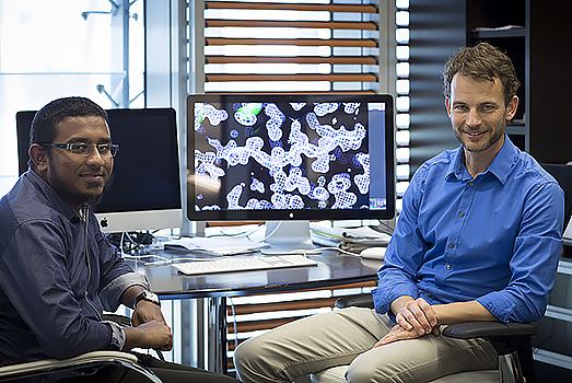
KAUST Associate Professor of Bioscience Stefan Arold (right) and postdoctoral fellow Umar Farook Shahul Hameed.
All organisms use protein-based communication networks to perceive and respond to changes in environmental conditions. These networks are also used in plants to sense and react to stress conditions, such as those resulting from drought or attack from pathogens. Increased demand, altered weather patterns and emergence of novel pathogens steadily augment stress on food crops. Increasing plant resilience is therefore key to assuring food security. If we understand in detail the interactions formed between proteins that communicate plant stress, we might be able to engineer more stress-tolerant plants. That is why a recent breakthrough by KAUST Associate Professor of Bioscience Stefan Arold’s group is of notable importance.
Making use of the powerful X-rays generated by the SOLEIL synchrotron in Paris, France, the KAUST group represented by postdoctoral fellow Umar Farook Shahul Hameed became the first to determine the atomic 3-D structure of a key protein for mediating plant stress signaling.
Arold explained: “My group is working on structural biology. In particular, we use X-ray crystallography, which is a method that allows the visualization of the 3-D atomic structure of proteins. The unique 3-D structure, which is made of tens or hundreds of thousands of atoms, enables the function of each protein, so unraveling the 3-D structure of proteins allows us to understand their function.”
Mapping the way towards transgenic plants
The work was carried out at the beamline PROXIMA II of the SOLEIL synchrotron, which is equipped with advanced microfocus optics that focalize the formidable X-ray beam on a surface as small as five micrometers. This setup was used to irradiate a tiny crystal formed by the plant enzyme. It took Hameed more than six months to optimize this fragile crystal of a size of less than one twentieth of a millimeter. The interactions of the X-rays with electrons making up each atom of each protein in the crystal result in a diffraction pattern. From this pattern, which consists of hundreds of thousands of data points, the 3-D protein structure can be obtained to atomic detail.
In order to capture the weak diffraction pattern from the tiny crystal, the team needed to use the most advanced detector for X-ray diffraction experiments, the EIGER 9M, which had just been installed at the PROXIMA II beamline. To obtain a full data set, the protein crystal is rotated by 0.1 degree, and for each rotation, a picture is recorded until a complete 180-degree view of the crystal is achieved. The EIGER 9M detector has the ability to capture a picture every 0.025 seconds, sensing every photon. That means it can record the 1,800 picture frames in 45 seconds.
“The EIGER 9M is not only extremely sensitive, but also extremely fast. In 45 seconds, Umar was able to obtain very beautiful data sets that allowed us to see every detail of the molecule,” said Arold. “We not only got the molecule but also a cofactor and the ligand, the protein that binds and activates it.”
Arold acknowledges that structure determination would not have been possible without the sensitivity and speed of the top-notch instrument. “We are very thankful to Drs. William Shepard, Martin Savko and Gavin Fox from the PROXIMA II beamline for their support and for allowing us to beta-test this fantastic setup. I’m glad we became the first to determine a novel structure with it,” he said.
Even though the protein is a tiny component of the plant, it is critical for its function and survival.
“Understanding the precise structure of the protein is significant because it plays a crucial role in the plants’ protective functions from stress tolerance to invading microorganisms,” Hameed noted. “When more information becomes available based on the structure, it might inspire mutants that can produce more resistant plants.”
Therefore, the vital step of characterizing the enzyme’s 3-D structure opens the door to understanding what role it plays in plant stress tolerance, and it will guide efforts to produce transgenic plants that might grow in desert conditions or in high salt concentration areas.
Delving deeper into the protein’s past
Although the enzyme is responsible for activating the cellular response to environmental stress in plants, similar proteins are also found in animals, including humans.
“Now that we’ve got the structure, it’s surprising how little it has changed over the period of 1 billion years since humans and plants diverged,” Arold added. “It’s such an important factor that nature hasn’t changed its overall structure ever since it arose in the unicellular ancestor of plants and animals.”
Although the protein, in its shape and composition, is highly conserved, it is used in many different contexts in different species. Understanding how similar proteins can selectively control different functions is an important challenge.
The enzyme that Arold and Hameed studied is derived from the plant Arabidopsis thaliana, a model organism used in plant biology. Yet there are in fact more than 20 similar proteins present in Arabidopsis. Now that the 3-D structure has been obtained for one of them, this facilitates the analysis of the 19 others. For the Arold group, follow-up work also includes studying the structural mechanisms by which the plant enzyme interacts with DNA and particular ions. These interactions have a central effect in modulating stress responses.
A collaborative effort: from atom to organism
This project to characterize the 3-D structure of the protein identified as key for plant stress and immunity is part of a collaborative effort with Professor of Plant Science Heribert Hirt’s plant science group at KAUST. Hirt’s group focuses on the fundamental biology of plants at the molecular and physiological level, their interactions with the physical (heat, salt and drought) and chemical environment, as well as their connections to other life forms.
“This type of interdisciplinary collaboration at KAUST is very powerful because we can link the information at the atomic level with the entire organism. Together the results should produce a beautiful and insightful story,” said Arold.
Related Links
- By Meres J. Weche, KAUST News

