
KAUST researchers develop technology that could make cancer diagnosis faster
Scientists at King Abdullah University of Science and Technology (KAUST) have developed a new stain-free imaging platform designed to analyze tissue samples more quickly and consistently, supporting future AI-assisted cancer diagnostics. The research forms part of KAUST’s Smart Health mission to develop technologies that improve cancer prevention, diagnosis, and treatment.
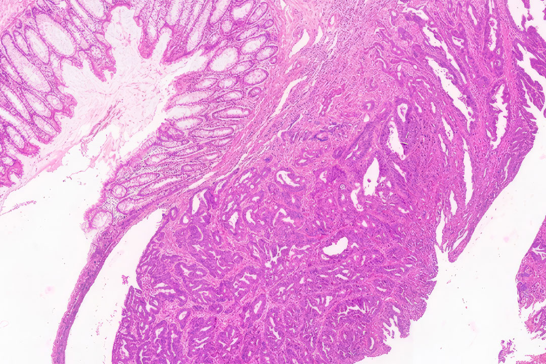
The platform was first validated using colorectal tissue samples, reflecting the importance of this disease area. Colorectal cancer remains a major health priority in Saudi Arabia, ranking among the most commonly diagnosed cancers in the Kingdom. Improvements in how these samples are analyzed could support earlier and more efficient diagnosis, helping to strengthen future care pathways.
Today, many pathology laboratories rely on chemical dyes to prepare tissue samples for microscopic examination. While widely used, this process can add time to diagnostic workflows and may vary depending on preparation methods and laboratory conditions.
The KAUST-led team has developed an alternative approach that uses engineered silicon slides to generate detailed structural color images directly from tissue samples, removing the need for conventional staining. The images can be reviewed by pathologists while also creating standardized data that could support future AI-assisted diagnosis.
In the study, the platform achieved a 99% agreement rate with conventional pathology assessments when analyzing colorectal tissue samples, meaning pathologists reached the same diagnostic conclusions in almost all cases while using a faster, stain-free imaging process.
The platform was evaluated using tissue samples from 120 patients, where researchers compared its performance against conventional pathology methods. The results showed strong agreement in how healthy and cancerous tissue features were identified, supporting further validation of the approach in clinical settings.
Because the method removes the need for chemical staining, the team also observed a reduction in preparation time compared with conventional workflows. Early results indicate the process could reduce sample preparation time by approximately 40-50%, while also improving consistency by removing variability linked to staining conditions.
"This research focuses on improving one of the most important steps in diagnosis: how tissue samples are prepared and reviewed," said Professor Qiaoqiang Gan, Professor of Material Science and Engineering at KAUST. "Traditional staining methods can be influenced by preparation steps, reagent quality, and laboratory conditions. By generating consistent digital images without dyes, we can reduce variability and create data that is more reliable for both clinical review and future AI-assisted analysis"
The platform has been developed with practical deployment in mind, and the research team is working to further validate the system and assess pathways for future clinical and commercial use.
The research brought together expertise from materials science, biomedical science and computing, reflecting KAUST’s interdisciplinary approach to diagnostic research. The team is now working with clinical partners, including King Faisal Specialist Hospital & Research Centre (KFSHRC) Madinah, to further evaluate the platform across broader healthcare settings in Saudi Arabia. By connecting discovery research with practical applications, KAUST provides an environment where new diagnostic technologies can be advanced toward real-world use.
The technology could also have future applications beyond colorectal cancer. In the study, researchers also tested breast, lung, and thyroid tissue samples, and the platform captured key histological features comparable to those on conventional-stained slides.
The study was published in Advanced Science.